A new, more comfortable way of detecting breast cancer, which could enable tumours to be identified at an earlier stage, has entered UK trials.
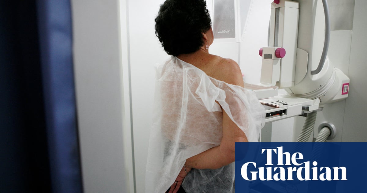
About one in eight British women will be diagnosed with breast cancer at some point in their lives. Although an X-ray test called a mammogram can spot tiny cancers in post-menopausal breasts, they are less effective in younger women whose breasts contain more dense, fibrous tissue and less fat, because cancers and fibrous tissue both show up as solid white areas on X-ray.
Mammograms can also miss cancers in postmenopausal women with relatively dense breast tissue, who are also at greater risk of developing breast cancer in the first place.
Here, women with a suspicious lump may be offered an ultrasound scan or a biopsy, and if the diagnosis is still uncertain they may be referred for so-called dynamic contrast enhanced magnetic resonance imaging (DCE-MRI), which identifies the growth of new blood vessels supplying tumours. However, these may not be visible in women with early-stage cancers, meaning they are given false reassurance.
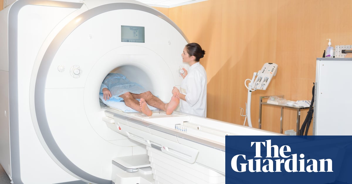
The new technique, called multiparametric MRI, was originally developed to evaluate liver diseases without the need for a painful biopsy, and is already in widespread used across Europe and the US.
Like conventional MRI, it uses strong magnetic fields and radiowaves to excite particles called protons in the tissue, using differences in the amount of time they take to settle to create a “map” of the various tissues in the breast. However, by combining images created by different MR pulses and sequences, multiparametric MRI enables an even more detailed map to be created.
“We believe if you differentiate the tissue, instead of looking at the blood vessels around the tumour, we should be able to spot not only tumours in dense breasts, but potentially tumours which aren’t seen on mammograms,” said Prof Sally Collins, a consultant obstetrician and medical lead for women’s health at the Oxford-based Perspectum Diagnostics, who herself recently underwent treatment for breast cancer.
“We’re also trying to improve the patient experience of being scanned. Mammograms are horrible – they basically squash your boob on this plate, which is undignified and uncomfortable – and MRIs are even worse because you have to lie face down with your boobs dangling in this coil and your arms above your head like Superman, and stay like that for ages,” Collins said.
“We’re trying to arrange it so that woman can be clothed and dignified and comfortable while being scanned, all of which is quite important for the patient journey through the diagnosis of cancer.”
Perspectum has ethical approval to recruit 1,030 women to the trial, including 10 with confirmed breast cancer and 30 to 40 healthy women who are currently being scanned, to test whether the technique can accurately map their breast tissue while they are lying on their back. The trial is expected to take about two years to complete.
“It is never going to replace routine mammogram screening for postmenopausal women, but we hope it will improve the diagnostic pathway for women with dense breasts or premenopausal women who are at very high risk of breast cancer, negating the need for multiple tests, which is what they’re going through at the moment,” Collins said.


